
Quantra® 2.2 Breast Density Assessment Software
Standardizing breast density analysis with machine learning, at the point of care.
Standardize Breast Density Analysis
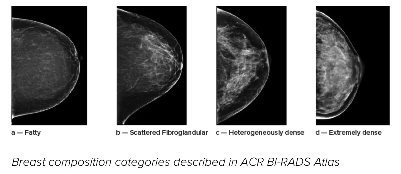
Higher breast tissue density increases a woman's risk for breast cancer1. Powered by machine learning, Quantra software provides accurate, unbiased breast density assessment by analyzing both 2D and tomosynthesis images for distribution and texture of tissue. Consistent with pattern and texture scoring guidance from the American College of Radiation (ACR) BI-RADS Atlas 5th Edition, Quantra software categorizes breast tissue into four breast density composition categories.
Better Risk Prediction
In addition to volume, pattern and texture of fibroglandular tissue may play just as an important role in mammographic cancer prediction.2-4 Both tumors and dense tissue show up as white on traditional mammogram images, impacting breast cancer screening3, making breast density a key metric in patient health. Quantra 2.2 software solution delivers the accurate information you need to achieve more consistent and reliable scoring and the confidence to design individualized patient screening plans.
Automated Assessment
Objective machine learning algorithm that assigns breast density category based on analysis of breast tissue texture and patterns.
Standardization
Overcome individual radiologist subjectivity in visual assessment and elevate the standard of care and standardization of reporting across the whole radiology practice.
Smoother Patient Pathway
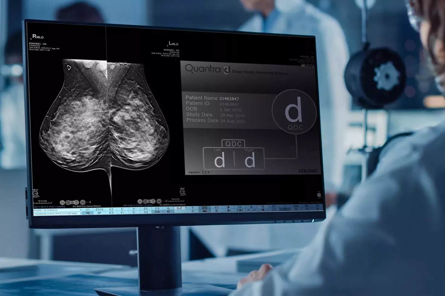
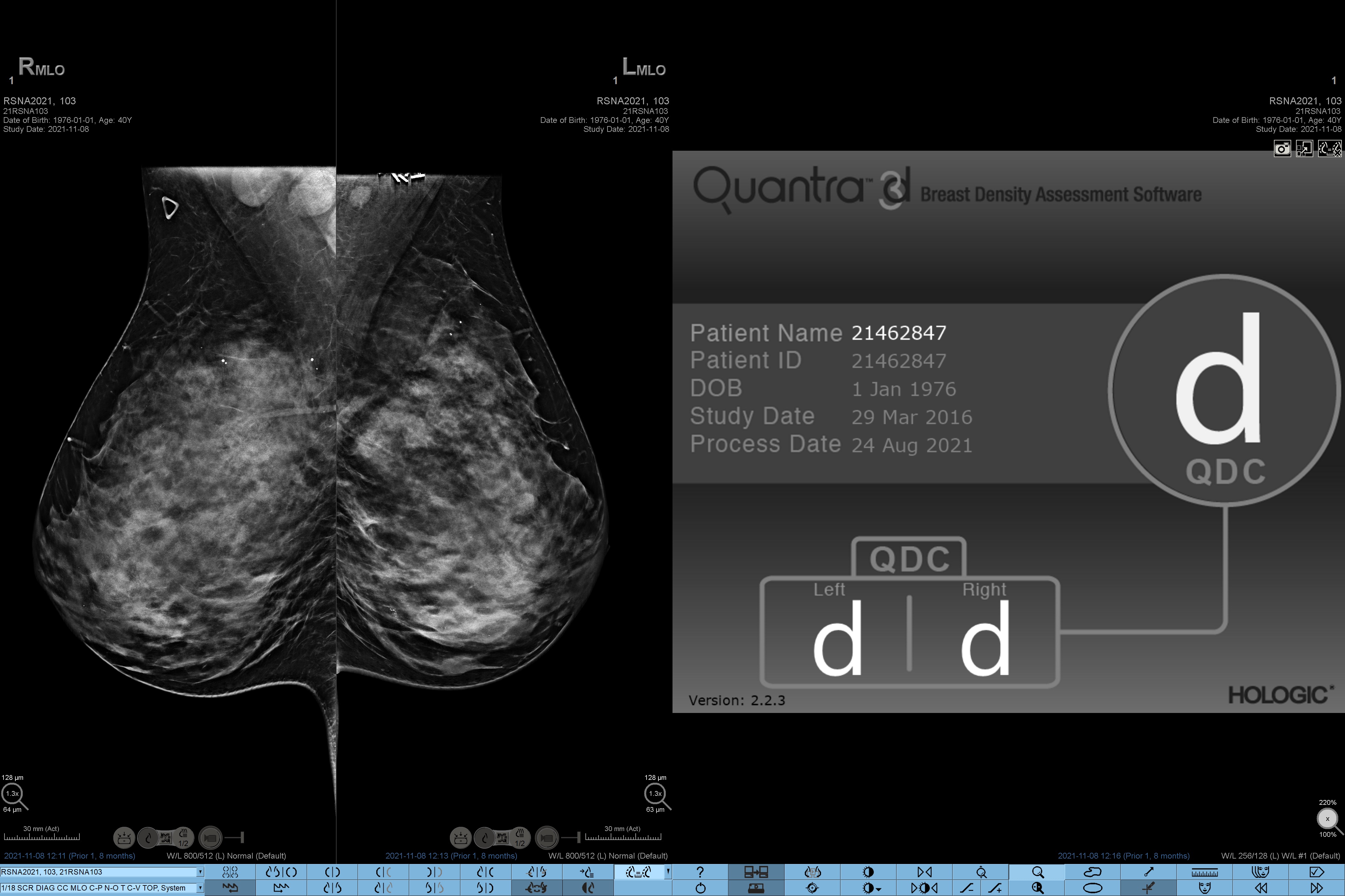
Displays density on the acquisition workstation in the exam room to facilitate patient management protocols for potential supplemental imaging at the point of care.
System Compatibility & Clinical Performance
Quantra 2.2 software solution allows you to seamlessly view results on SecurView® and other diagnostic workstations and mammography reporting systems. It can also be used as an automatic input for risk assessment software that utilizes breast density scores, such as the Tyrer-Cuzick model.
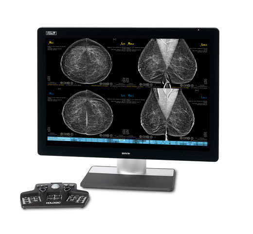
View breast density scores on the 3Dimensions™ and Selenia Dimensions® systems’ acquisition workstation to streamline workflows for supplemental screening, and eliminate the need for a separate server.
Risk Categories8
Highest risk category: left breast D, right breast D
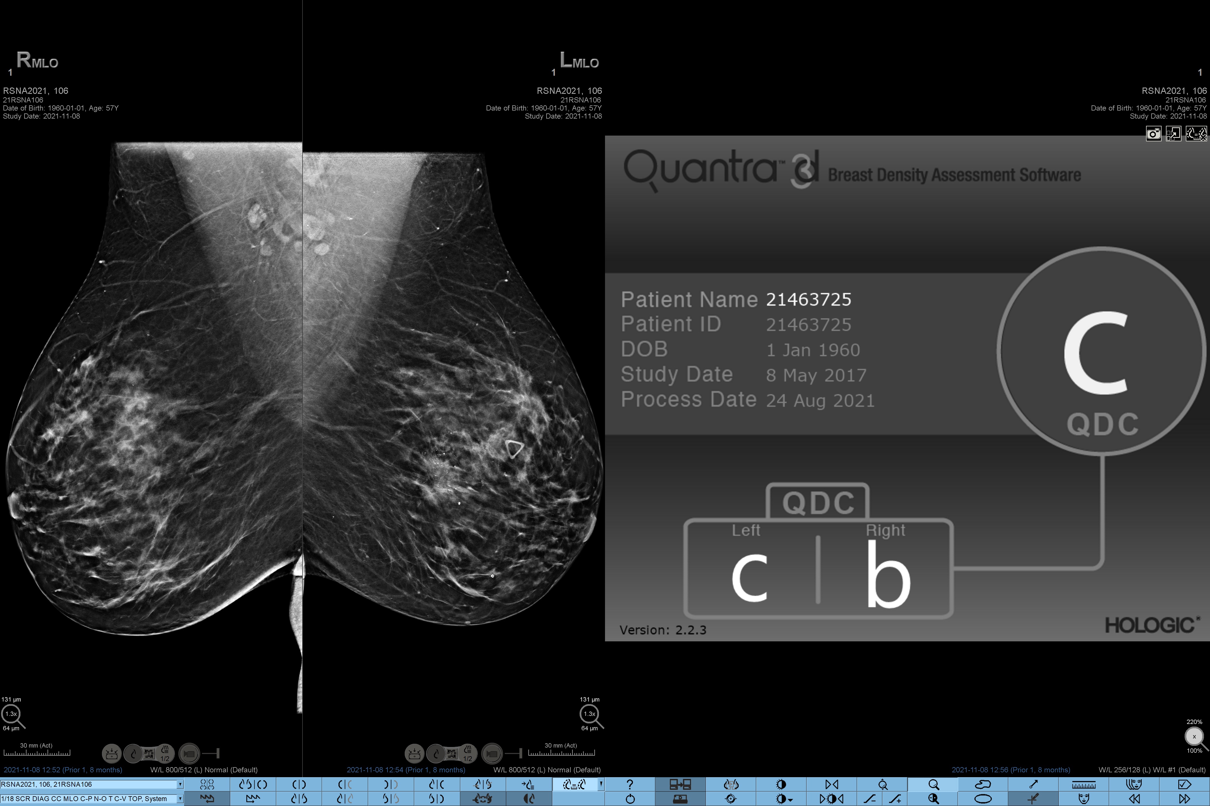
Varying risk category: left breast C, right breast B

Varying risk category: left breast A, right breast B
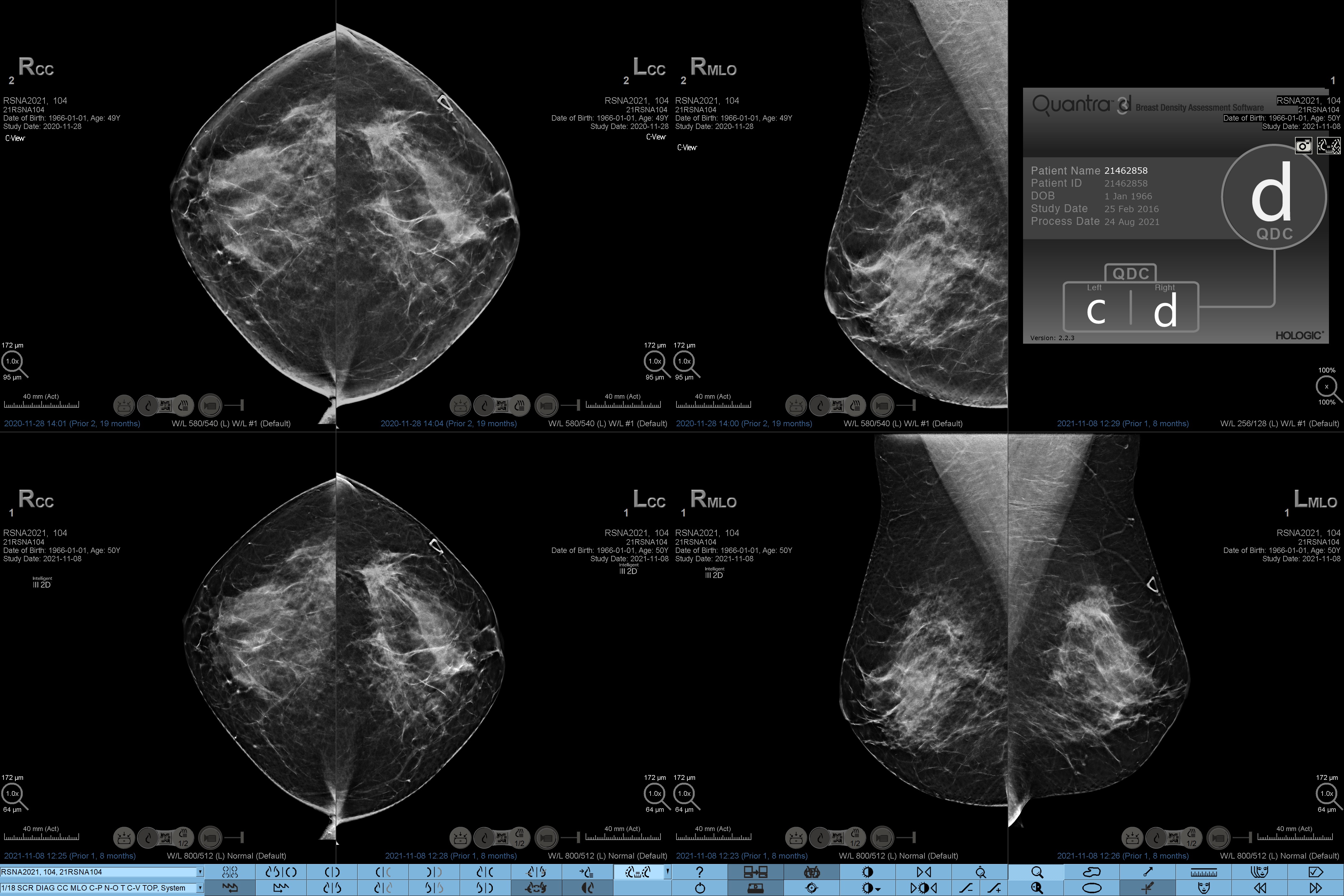
Varying risk category: left breast C, right breast D
Evidence. Insight. Collaboration.
Our education portal improves patient care through excellence in education, communication of clinical and scientific evidence, and partnerships with the healthcare community.