
Overview
Clinical Images
Documents
Training
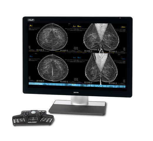
One powerful platform.Many imaging possibilities.
Create comprehensive diagnostic studies that reveal exceptional clinical detail and sensitivity with contrast-enhanced mammography (CEM) imaging.
Turn the Invisible Into the Visible
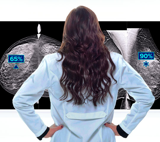
Clinical Performance
Increase diagnostic confidence to help guide the clinical pathway. Studies show contrast-enhanced mammography provides equivalent diagnostic performance to contrast breast MRI.1,2
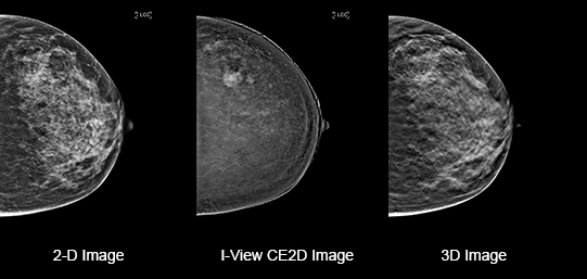
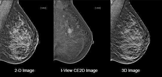
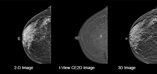
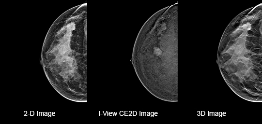
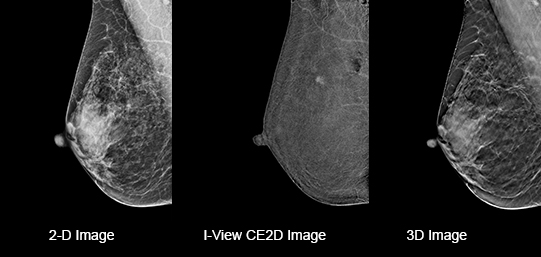
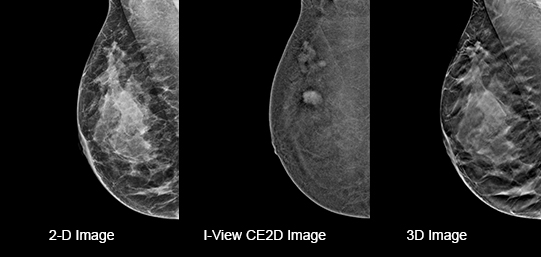
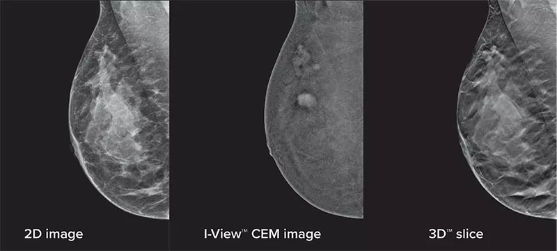
Eliminates Structural Background
Acquire a pair of high- and low-energy images in rapid succession. This allows for regions of abnormal blood flow to be displayed by subtracting the background breast parenchyma.
Patient Preference
Patients greatly prefer contrast-enhanced mammography over breast MRI for reasons such as faster procedure time, greater comfort, lower anxiety, and lower noise level.3
Expand Diagnostic Capabilities
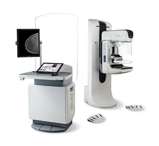
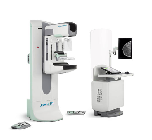
I-View 2.0 Software is a simple upgrade to any Selenia® Dimensions or 3Dimensions mammography system. Offering CEM exams expands your capabilities using existing mammography equipment.
More Diagnostic Information
Contrast-enhanced mammography enhances visualization and may uncover hidden abnormalities – a crucial factor in reducing missed and or underdiagnosed cancers.
The future of contrast-enhanced mammography is bright.
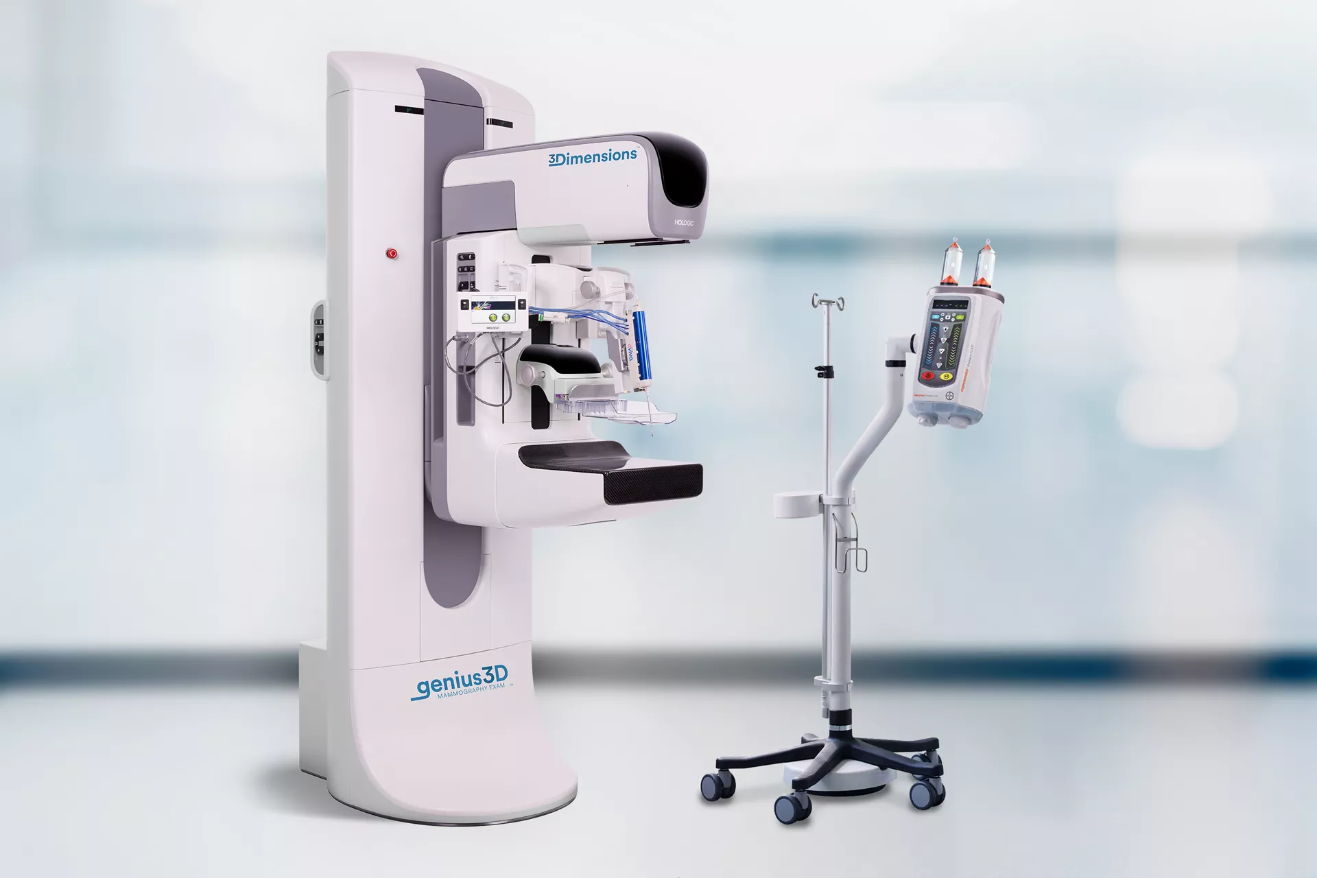
I-View® 2.0 CEM Software together with MEDRAD® Stellant FLEX CT Injection System with Certegra® Workstation.
Hologic and Bayer have come together to deliver a coordinated solution for Contrast- Enhanced Mammography with a robust network of supporting expertise. Together we can make this modality accessible to health care providers and their patients, by championing educational resources, offering streamlined acquisition pathways, and providing ongoing supportive clinical expertise - for simple and successful implementation in facilities.
![]()
Create a better patient experience
CEM is a highly sensitive and relatively low-cost breast imaging modality compared to breast MRI.4
A faster procedure
8-10 minutes5 for CEM vs. 45 minutes4 for breast MRI.
More comfortable positioning
Avoids uncomfortable prone position and anxiety associated with the noisy and enclosed MRI tube.3
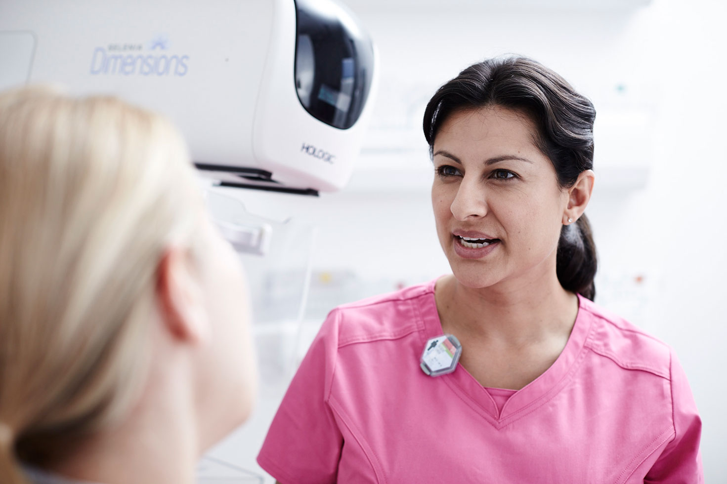
Familiar exam
Same room and equipment as the diagnostic mammogram.
Important safety information:
- Patients can have adverse reactions to contrast agents. Refer to the Instructions for Use for the contrast agent for complete information.
- Contrast biopsy uses contrast agents that are injected intravenously. Allergic reactions can occur.
1. Chou C, Lewin J, Chiang C et al. "Clinical Evaluation of Contrast-Enhanced Digital Mammography and Contrast Enhanced Tomosynthesis-Comparison to Contrast-Enhanced Breast MRI" Eur J Radiol. 2015 Dec; 84(12):2501-8. [Epub 2015 Oct 1]. 2. Jochelson M, Dershaw D, Sung J, et al., Bilateral contrast-enhanced dual-energy digital mammography: feasibility and comparison with conventional digital mammography and MR imaging in women with known breast carcinoma, Radiology 266 (3) (2013) 743–751 3. Hobbes M, Taylor D, Buzynski S et al. “Contrast-enhanced spectral mammography (CESM) and contrast enhanced MRI (CEMRI): Patient preference and tolerance” J Med Imaging Radiat Oncol. 2015 Jun;59(3):300-5. [Epub 2015 Apr 21]. 4. https://www.radiologyinfo.org/en/info.cfm?pg=breastmr= 5. Patel B, Gray R, Pockaj B. Potential Cost Savings of Contrast-Enhanced Digital Mammography. AJR 2017;208:W231-W237. 6. Covington MF. Contrast-Enhanced Mammography Implementation, Performance, and Use for Supplemental Breast Cancer Screening. Radiol Clin North Am. 2021 Jan;59(1):113-128. doi: 10.1016/j.rcl.2020.08.006. Epub 2020 Oct 29. PMID: 33222993.
Bayer and the Bayer Cross are trademarks owned by and/or registered to Bayer in the U.S. and/or other countries.
Clinical Images