

Contrast-Enhanced Mammography Explained: A Conversation with Hologic Scientist Wei Wang
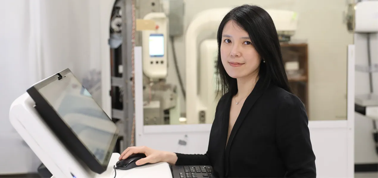
Dr. Wei Wang.
Nearly half of women have dense breasts, which can make tumors harder to see on mammograms.1 Contrast-enhanced mammography, or CEM, is an imaging option that may help radiologists detect and evaluate breast cancer by highlighting “leaky” blood vessels associated with tumors.
What is contrast-enhanced mammography?
During a CEM exam, an iodine‑based contrast agent is injected into the patient’s bloodstream before imaging. Areas with abnormal blood vessel growth may take up more contrast than surrounding tissue, helping to highlight suspicious areas.
The technology then captures low- and high-energy X-ray images and processes them to produce both a standard mammogram and a recombined image.
“When we combine the images, we suppress the background tissue and highlight the iodine uptake,” says Wei Wang, Ph.D., Associate Director, R&D Innovation at Hologic. “If an area ‘lights up,' it may indicate the kind of abnormal vascular activity often associated with cancer.”
Contrast-enhanced mammography may be used to:2,3
- Support supplemental screening in women with dense breast tissue or who are at higher risk.*
- Evaluate and clarify inconclusive findings from standard screening, including masses and calcifications.
- Help assess the extent of disease, support treatment planning, monitor response to treatment and evaluate for recurrence.
- Provide an option when MRI is not feasible or practical, including for patients with implanted devices, claustrophobia, or limited access.
Quote from Wei Wang, Ph.D.
“For a long time, CEM was viewed as a supplemental breast imaging tool used in select situations. What’s changing now is the recognition that functional information isn’t just a supplement to structural imaging; it’s another important dimension of information for radiologists. As clinicians and researchers build more experience with this technology, we’re seeing it integrated more thoughtfully into imaging workflows.”
While CEM has its benefits, as with any procedure or treatment, it can have risks, such as allergic reaction to the contrast used in the exam. Most reactions are mild but can be severe in rare cases.
Helping reveal cancers in dense breast tissue
Dense breasts can make mammograms harder to read. That's because dense breast tissue looks white on a mammogram. So do some abnormal changes, like calcifications and tumors. Fatty tissue, by contrast, looks dark. This can make it harder to spot potential problems in dense breasts.4
“Even if the tissue is dense, cancers may stand out in CEM because they often develop abnormal blood vessels,” Dr. Wang explained. “Contrast imaging can help reveal that biology, so cancers that might be hidden on a regular mammogram can become visible.”
For women with dense breasts, clinicians may consider additional imaging based on breast density, personal risk, symptoms and other factors.
An alternative to MRI
Breast MRI is widely used to visualize blood flow in breast tissue, but exams can be expensive and difficult to schedule in some settings.5 Some patients also cannot tolerate MRI because of claustrophobia or implanted devices.
A CEM exam typically takes 6-10 minutes compared with 45 min with a breast MRI.6 The exam can also be performed on compatible mammography systems with additional hardware and software, allowing facilities to add the capability without the infrastructure investment of a separate imaging modality.
“MRI is powerful, but it isn’t always available at every medical center or practical for every patient,” Dr. Wang said. “Contrast-enhanced mammography can provide clinically meaningful functional information in a more familiar breast imaging environment, which can help expand access — especially in community settings.”
New frontiers
Clinicians and researchers are exploring ways to extract more information from CEM beyond what the eye can see alone. Today, radiologists primarily assess whether and where tissue enhances, or “lights up." Ongoing work in the field is examining how image analysis techniques may support interpretation and clinical decision-making.
These efforts broadly include approaches that assess patterns within images as well as artificial intelligence (AI) methods designed to support cancer detection and lesion characterization.
Researchers are also investigating how imaging data, in combination with other clinical information, might contribute to a more comprehensive understanding of breast health over time. These areas remain under evaluation, and their potential clinical applications continue to evolve.
Quote from Wei Wang, Ph.D.
“The question is no longer just whether something enhances,” Dr. Wang said. “The bigger opportunity is how to extract more meaningful information from the image in a way that supports radiologists and improves decision-making for patients.”
Contrast-enhanced mammography has been used clinically for more than a decade, and its evidence base has grown substantially. Dr. Wang believes the field is now entering a more expansive phase.
“The questions we’re now beginning to ask with CEM — about tumor biology, risk and treatment response — go beyond what this technology was initially used for,” Dr. Wang said. “That’s what makes this such an exciting time. As CEM evolves, the goal remains the same: better information for clinicians and better decisions for patients.”
Learn more about Hologic’s contrast-enhanced mammography and how we continue to shape the future of the field.
*Emerging evidence and guidelines recognize contrast-enhanced mammography as a potential option for selected patients, although optimal selection criteria continue to evolve.
- Dense Breasts: Answers to Commonly Asked Questions. (2025). In Cancer.gov. https://www.cancer.gov/types/breast/screening/dense-breasts
- Contrast-enhanced spectral mammography (CESM) and contrast enhanced MRI (CEMRI): Patient preference and tolerance” J Med Imaging Radiat Oncol. 2015 Jun;59(3):300-5. [Epub 2015 Apr 21].
- CDC. About Dense Breasts. 2024. Accessed July 28, 2026. https://www.cdc.gov/breast-cancer/about/dense-breasts.html.
- Weaver, O. (2024). What is a contrast-enhanced mammogram? In MD Anderson Cancer Center. https://www.mdanderson.org/cancerwise/what-is-a-contrast-enhanced-mammogram.h00-159701490.html.
- Patel B, Gray R, Pockaj B. Potential Cost Savings of Contrast-Enhanced Digital Mammography. AJR 2017;208:W231-W237
- The Principles of Contrast Mammography. Andrew Smith, Ph.D. (WP-00084 Rev02).